COVID: 3D video of how the coronavirus affects the lungs
American researchers at George Washington University Hospital have created a 3D video of lungs infected by the coronavirus, with frightening results.
They say a picture is worth a thousand words, but what about a 3D video? Every day, scientists and doctors discover new information regarding the symptoms of COVID-19. Now for the first time, the devastation that the coronavirus does to the lungs can be seen.
Visualisation of the infection
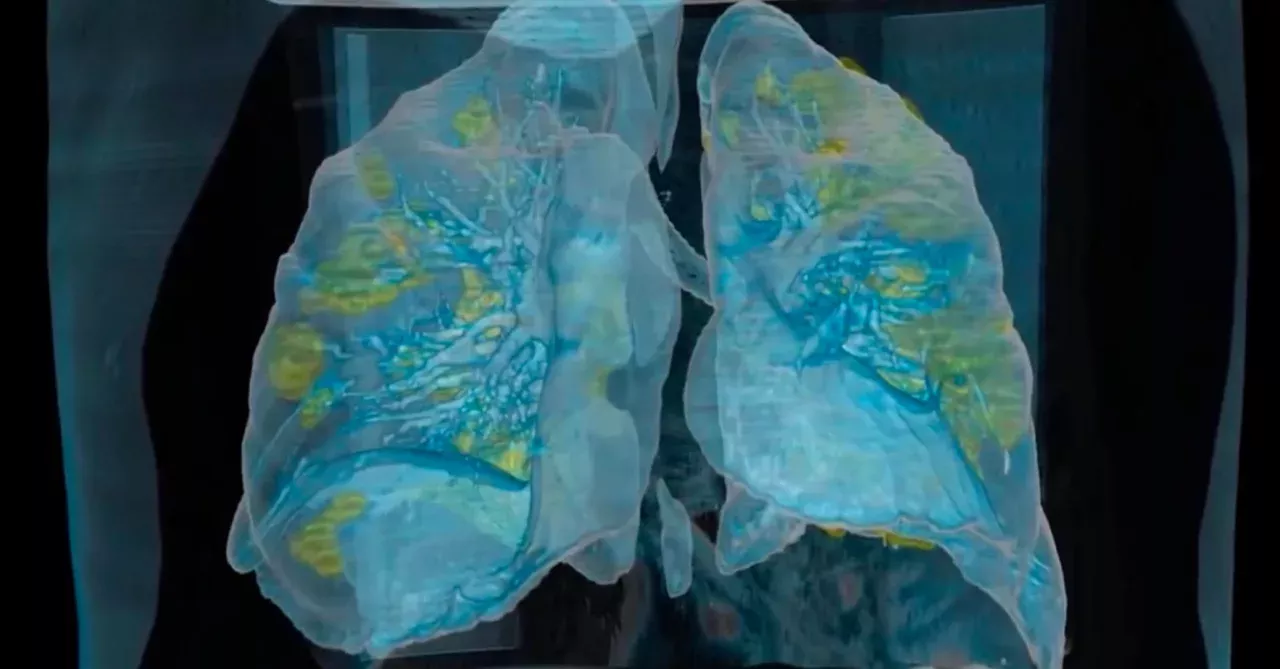
A 3D video was created by American scientists at George Washington University Hospital. The lungs that can be seen are those of a 50-year-old coronavirus patient, who was admitted to intensive care and placed on a ventilator.
Dr Keith Mortman, head of the Department of Thoracic Surgery at George Washington Hospital, was interviewed by numerous American media outlets:
There is such a contrast between the lung infected with the disease, and the adjacent healthy lung tissue, that you don't even need to be a doctor to understand these images, and how serious the coronavirus can be
In the video, healthy lung tissue is shown in blue, while areas infected with COVID-19 appear as a yellowish colour. The infection can, therefore, be seen throughout the lungs.
How the video was developed
The ‘Surgical Theatre’ software was used to create the video. This tool allows you to explore parts of the body as closely as possible, and to transform medical data, such as medical imaging, into virtual reality images.